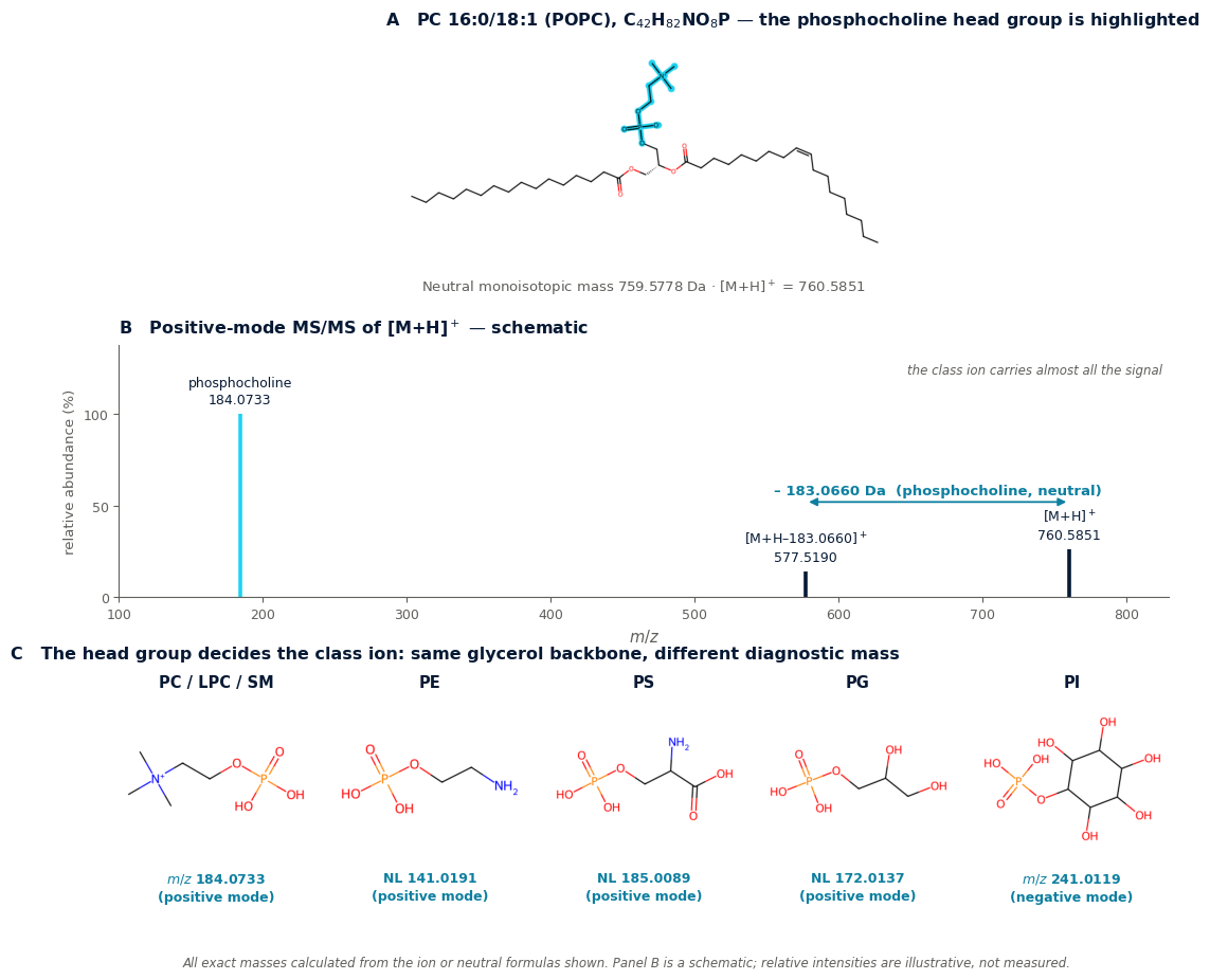
Lipids are the clearest case in all of LC-MS of a compound class announcing itself. Every phosphatidylcholine, whatever its fatty acyl chains, gives m/z 184.0733. Every phosphatidylethanolamine loses 141.0191 Da. The head group decides the diagnostic mass; the chains decide only where the precursor sits. This is a reference page for those class signatures.
The principle. Lipid class-selective fragments are defined by the property that all molecules of a class yield the same fragment. In positive mode this means a class ion (PC: 184.0733) or a class neutral loss (PE: 141.0191). Screen with a precursor-ion scan for the ion, or a neutral-loss scan for the loss, and you find the whole class at once.
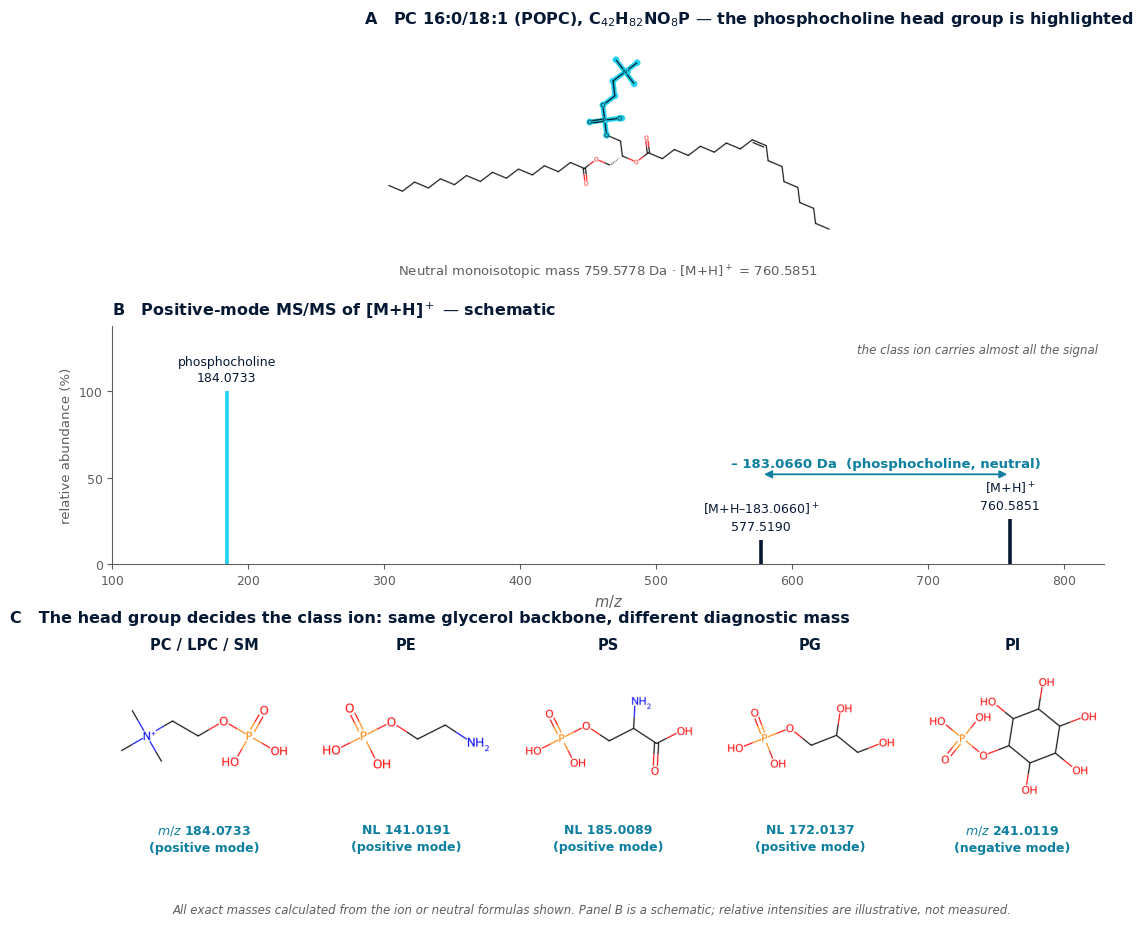
Class-diagnostic ions and losses at a glance
| Lipid class | Signature | Exact value | Formula | Mode |
|---|---|---|---|---|
| PC, LPC, PC O-, SM | Product ion | 184.0733 | C5H15NO4P+ (phosphocholine) | positive |
| PE, PE O- | Neutral loss | 141.0191 | C2H8NO4P (phosphoethanolamine) | positive |
| PS | Neutral loss | 185.0089 | C3H8NO6P (phosphoserine) | positive |
| PS | Neutral loss | 87.0320 | C3H5NO2 (serine residue) | negative |
| PG | Neutral loss | 172.0137 | C3H9O6P (glycerophosphate) | positive |
| PA | Neutral loss | 97.9769 | H3PO4 | positive |
| PI | Product ion | 241.0119 | C6H10O8P− (inositol phosphate) | negative |
| Ceramide, SM | Product ion | 264.2686 | C18H34N+ (d18:1 sphingoid backbone) | positive |
| Cholesteryl ester | Product ion | 369.3516 | C27H45+ (cholestadienyl cation) | positive |
| TAG, DAG, PA, steryl ester | Neutral loss | 17.0265 | NH3 from [M+NH4]+, then the acyl chains | positive |
| PC, LPC, PC O-, SM | Neutral loss | 60.0211 | C2H4O2 (methyl formate, from formate adducts) | negative |
| PC | Product ion | 168.0426 | reported value; formula not assigned here | negative |
The concept has a formal definition. Lipid class-selective fragments are characterised by the property that all molecules belonging to the same class yield the same fragment — m/z 184.0733 from all protonated LPC, PC, PC O- and SM species, and neutral loss 141.0191 from protonated and sodiated PE and ether PE (Peer-reviewed evidence).
Screening scans built on these values
A published HILIC lipidomics protocol separates and detects phospholipid and sphingolipid classes using precursor-ion scans for m/z 184, 241 and 264, and neutral-loss scans for 141, 172, 185 and 98 — corresponding to PC, PI and ceramide, and to PE, PG, PS and PA respectively (Peer-reviewed evidence).
| Scan type | Set to | Finds |
|---|---|---|
| Precursor ion | 184.0733 | PC, LPC, PC O-, SM |
| Precursor ion | 241.0119 | PI |
| Precursor ion | 264.2686 | Ceramides, sphingolipids with d18:1 |
| Neutral loss | 141.0191 | PE, LPE |
| Neutral loss | 172.0137 | PG |
| Neutral loss | 185.0089 | PS |
| Neutral loss | 97.9769 | PA |
Identifying the chains as well as the class
The class ion identifies the head group and nothing else. To get the acyl chains you need the complementary fragments:
- Positive mode, glycerophospholipids: the loss of the head group leaves a diacylglycerol-type ion — for PC 16:0/18:1, [M+H−183.0660]+ at m/z 577.5190. Further losses of each acyl chain as a ketene or a free fatty acid then reveal the individual chains.
- Negative mode: the carboxylate anions of the fatty acyl chains appear directly, which is why negative mode is preferred for chain assignment.
- Neutral lipids: triacylglycerols, diacylglycerols and steryl esters ionise as ammonium adducts. CID gives the loss of ammonia (17.0265 Da) combined with charge-remote loss of the fatty acyl moieties as fatty acids (Peer-reviewed evidence).
- Cholesteryl esters: the cholesteryl motif at m/z 369.3516 is the key diagnostic fragment (Peer-reviewed evidence).
What the class ion cannot tell you
A precursor-ion scan of m/z 184 is the standard approach for PC and SM, but it cannot distinguish ether-linked PC species from diacyl species, and it cannot identify the sphingoid base in SM (Peer-reviewed evidence). Likewise, PE annotation — especially of plasmalogens — should not rest on the head-group-specific neutral loss of 141 alone: such scans identify the class but do not provide species-level analysis (Peer-reviewed evidence).
Practical consequence: report the class from the class ion, and the species only when the chain fragments support it. Sum composition such as “PC 34:1” is a weaker and more honest claim than “PC 16:0/18:1” unless the individual chains were observed.
Two further limits worth stating. The 264.2686 ion identifies a d18:1 sphingoid backbone specifically — other backbones give other masses, so a negative result is not evidence against a sphingolipid. And on Orbitrap systems, many lipid classes fragment under HCD in a way that is characteristic but structurally uninformative, which is why staged activation is used when chain-level detail matters (Peer-reviewed evidence).
Practical notes
- Choose the adduct deliberately. Ammonium salts or weakly acidic modifiers give protonated choline-containing phospholipids; formate gives formate adducts with their own negative-mode losses. The adduct changes the fragmentation, not just the precursor mass. (Practical starting point)
- Run both polarities where possible: positive for class assignment, negative for chain assignment.
- Use the exact mass, not 184. Nominal-mass class filters catch unrelated ions; 184.0733 does not.
- Separate the classes chromatographically — HILIC groups lipids by head group, which pairs naturally with class-specific scans.
References
- Proposal for a common nomenclature for fragment ions in mass spectra of lipids. PLOS ONE 2017;12(11):e0188394 — source for the definition of lipid class-selective fragments, m/z 184.0733 for LPC/PC/PC O-/SM, neutral loss 141.0191 for PE and PE O-, the 60.0211 Da methyl formate loss from formate adducts, and the 17.0266 Da ammonia loss from ammonium adducts of TAG, DAG, PA and steryl esters. doi:10.1371/journal.pone.0188394 — open access; also at PMC5697860.
- Lipidomics of human tissues using ion mobility MS — LIPID MAPS protocol. Source for the precursor-ion scans at m/z 184, 241 and 264 and the neutral-loss scans at 141, 172, 185 and 98, and their assignment to PC, PI, ceramide, PE, PG, PS and PA. lipidmaps.org (PDF)
- Shorthand notation for lipid structures derived from mass spectrometry. Journal of Lipid Research — source for the limits of the m/z 184 precursor scan (ether versus diacyl PC, sphingoid base in SM) and of NL 141 for PE annotation. PMC3646453
- Improved Structural Characterization of Glycerophospholipids and Sphingomyelins with Real-Time Library Searching — source for the m/z 264.268 sphingoid backbone ion and for the observation that many lipid classes fragment characteristically but uninformatively under HCD. PMC10840458
- Comprehensive LC-MSE lipidomic analysis using a shotgun approach — source for the cholesteryl motif at m/z 369.3516 as the key diagnostic fragment of cholesteryl esters and for the ammonium-adduct behaviour of neutral lipids. PDF
- LC-ESI-HRMS lipidomics of phospholipids. Analytical and Bioanalytical Chemistry 2024 — source for the use of m/z 184.0733 (positive) and 168.0426 (negative) as class-confirming criteria in an identification workflow. doi:10.1007/s00216-023-05080-0
Exact masses in the tables were calculated from the ion or neutral formulas given, using standard atomic masses and correcting for the electron mass, and agree with the published values. The structures in Figure 1 were generated from SMILES and their molecular formulas verified computationally. References 1–3 and 5 predate this series’ usual reference window; they are cited for class fragmentation behaviour, which is not time-dependent.
Leave a Reply