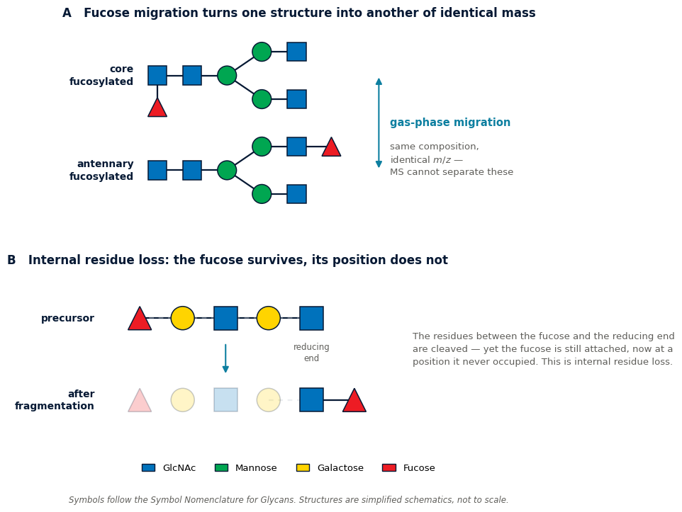
Every structural interpretation of an MS/MS spectrum rests on one unspoken assumption: that the fragments reflect the connectivity of the molecule that entered the instrument. For a large and well-documented class of compounds that assumption is wrong. Groups migrate inside the ion — before it breaks, and sometimes before it is even activated — and the resulting fragments look exactly like evidence for a structure that was never there.
Quick answer. In the gas phase, substituents can move from one position to another within an ion. The best-documented case is fucose migration in glycans, but the same has been reported for mannose, rhamnose, xylose, GlcNAc and sulfate groups, and analogously as sequence scrambling in peptides. Crucially, migration is not simply a consequence of harsh collision energy: gas-phase IR spectroscopy has shown fucose migration in intact protonated ions with no CID at all, which points to a low energy barrier and makes it a universal risk. The practical defences are negative-ion mode, derivatisation that traps the mobile proton, ion mobility, and refusing to assign a position from a single fragment.
The mechanism: a mobile proton and a low barrier
Collision-induced dissociation deposits energy into an ion, and that energy is redistributed over the whole structure before anything breaks. If a rearrangement pathway has a lower barrier than the bond you intended to cleave, the rearrangement happens first.
For protonated glycans the catalyst is the proton itself. A 2025 review in Angewandte Chemie by Geue and co-workers summarises the mechanistic picture: charge migration of the mobile proton is thought to catalyse the rearrangement of individual fucose residues onto spatially adjacent sites [1] (Peer-reviewed evidence). The proton wanders, the sugar follows.
The consequence is uncomfortable. What you measure is the mass of a fragment; what you infer is where that fragment sat. Migration breaks the link between the two.
Fucose migration: the best-documented case
Internal residue loss
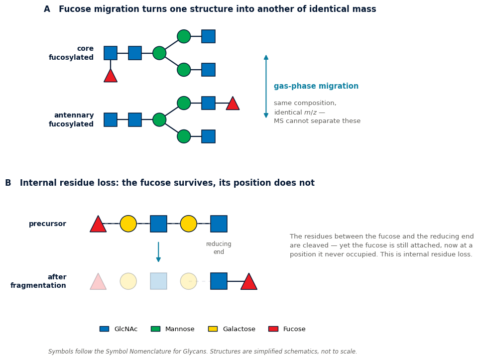
The phenomenon was first described in the mid-1990s by Kováčik and colleagues and named internal residue loss. In fucose-containing oligosaccharides, the sugars between a fucose near the non-reducing end and the reducing end are cleaved away — yet the fucose ends up attached directly to the reducing-end sugar, a position it never occupied [2] (Peer-reviewed evidence, as reported in the 2026 study cited below).
The resulting ion has a perfectly sensible mass and a perfectly wrong structure. As one mechanistic study of the migration destinations puts it, such rearrangements can be mistaken for diagnostic fragments and lead to false assignment
of the fucose position on the glycan core [4].
It happens without CID
This is the finding that changed how the problem is understood. Using gas-phase infrared spectroscopy, researchers showed that fucose migration also occurs in intact protonated oligosaccharide ions in the absence of CID. The Angewandte Chemie review states the implication directly — that fucose migration is a universal phenomenon in MS due to a low energy barrier
[1] (Peer-reviewed evidence).
A 2026 mechanistic study in Communications Chemistry reaches the same conclusion from a different angle. Having established that the process requires a mobile proton, the authors conclude that the rearrangement is not induced by the CID-induced fragmentation
but is most likely triggered by in-source activation during nanoelectrospray [2] (Peer-reviewed evidence).
Turning down the collision energy does not solve this. If the migration has already happened in the source, a gentler MS/MS simply fragments an ion that was rearranged before you touched it. Lowering the energy is worth doing, but it is not a fix.
Where the fucose goes
Migration is not random. Work on the destination of the migrating residue found that fucosyl groups move not to hydroxyls but to acetamido moieties of N-acetylneuraminic acid and N-acetylglucosamine residues, and to nucleophilic sites on an anomeric tag, producing specific isomeric fragment ions. Because the products are defined rather than arbitrary, they can be recognised — ion mobility gives them distinguishable arrival time distributions, which in turn allows the true fucose position to be determined [4] (Peer-reviewed evidence; 2022 publication, outside this series’ usual reference window).
A further refinement reported by Moge and colleagues is intra-residue migration: a fucose at O3 or O4 of a GlcNAc residue can migrate to O6 of the same residue — additional evidence for how low the barrier is [1] (Peer-reviewed evidence).
A concrete example
For the Lewisy antigen analysed as [M+H]+, the main fragment at m/z 530 is itself the product of fucose migration [1] (Peer-reviewed evidence). Not a minor side peak — the dominant signal in the spectrum. Gas-phase IR spectra were needed to establish this, by comparing the fragment against intact trisaccharide standards of the same nominal mass.
This matters for anyone reading the deoxyhexose loss of 146.0579 Da described in our article on glycoside fragmentation. The loss reliably tells you a deoxyhexose was present. It does not reliably tell you where it was attached.
Other groups that migrate
| Migrating group | Observed in | Consequence for interpretation |
|---|---|---|
| Fucose (deoxyhexose) | Oligosaccharides, blood-group antigens, N-glycans | False assignment of fucose position; migration products can dominate |
| Mannose | Oligosaccharides | Altered apparent branching |
| Rhamnose, xylose | Plant and microbial glycans | Same class of positional error |
| GlcNAc (reducing end) | N-glycans | Migrates onto the mannose antennae, producing an apparent loss of a core GlcNAc |
| Sulfate | Glycosaminoglycans | Sulfation site cannot be read directly from the fragment |
| Whole peptide segments | Peptide b ions | Sequence scrambling — non-sequence ions that look like sequence ions |
The GlcNAc case, reported by Wuhrer and colleagues, deserves attention because the outcome is deceptive in a specific way: a residue moves onto a branch, and the spectrum then shows what looks like the loss of a core residue. Nothing was lost; something moved [2] (Peer-reviewed evidence).
The peptide parallel: sequence scrambling
Proteomics met this problem two decades ago and largely solved it, which is why the glycomics literature explicitly frames fucose migration as the same kind of challenge that peptide sequence scrambling once posed [1] (Peer-reviewed evidence).
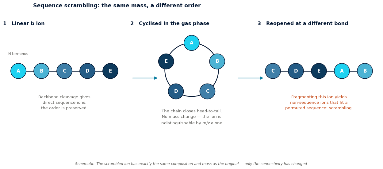
The mechanism is cyclisation. A b ion can close into a ring in the gas phase and then reopen at a different point. Fragmenting that reopened ion yields non-direct sequence ions — fragments that correspond to a permuted version of the original sequence. They score like real sequence ions in a database search, and in extreme cases can produce a false peptide identification [5] (Peer-reviewed evidence; 2008–2014 publications, outside this series’ usual reference window).
Two lessons from that history transfer directly:
- Derivatisation helps. Raising the gas-phase basicity of the N-terminal residue — converting lysine to homoarginine — reduces scrambling by holding the proton in place [5]. The glycan field applies the same idea, using fluorescent labels or other tags to trap the proton in positive-ion mode [1] (Peer-reviewed evidence).
- The problem is worse deeper in the tree. Scrambling was found to be a minor issue in direct MS2 of intact species but significant when large b ions were further fragmented in MS3 [5]. Each additional activation step is another chance to rearrange.
Small molecules are not exempt
Rearrangement is not confined to biopolymers. A 2025 study by Muste and co-workers documented macrocyclic rearrangement ion fragmentation in glutathione conjugates of cyclobutane-containing covalent kinase inhibitors — a case where the fragmentation of a drug metabolite proceeds through a rearranged cyclic intermediate [3] (Peer-reviewed evidence).
Anyone assigning metabolite structures from fragment shifts, as described in our article on the +16 hydroxylation shift, should treat this as a caution: the shift tells you an oxygen was added; the fragment carrying that shift tells you where it was added only if nothing moved.
Not everything is scrambled
An honest counterweight. Some structural information demonstrably survives fragmentation intact. Work by Compagnon and co-workers on the ring-size memory of galactose-containing oligosaccharides showed that precursor ions containing galactofuranose (five-membered ring) and galactopyranose (six-membered ring) retain the same ring size in their fragments — a result the authors themselves found surprising given the extent of rearrangement otherwise seen [1] (Peer-reviewed evidence).
Migration is a real and widespread hazard, not a reason to distrust every spectrum. The task is knowing which features are load-bearing and which are not.
Troubleshooting table
| Observation | Possible migration effect | Diagnostic check | Corrective action |
|---|---|---|---|
| Fragment supports a fucose position that contradicts biology or the enzyme used | Fucose migration | Compare with negative-ion CID of the same species | Assign composition, not position, from positive-mode CID |
| Apparent loss of a core residue that should be conserved | Residue migrated onto a branch | Check whether total composition is unchanged | Re-interpret as rearrangement, not cleavage |
| Two samples known to differ isomerically give near-identical spectra | Both converged on the same rearranged ion | Add ion mobility; compare arrival time distributions | Separate isomers before or after the mass measurement |
| Dominant fragment does not fit any expected cleavage | The migration product is the base peak | Compare against an authentic standard of that m/z | Do not build the assignment on it |
| MS3 spectrum contradicts the MS2 interpretation | Rearrangement during the extra activation step | Repeat with a different activation method | Weight MS2 evidence more heavily |
| Sulfation site differs between replicates | Sulfate migration | Compare fragment sets across collision energies | Report sulfation degree, not position, without orthogonal support |
| Peptide identification with an odd ion series | Sequence scrambling via b-ion cyclisation | Check whether unmatched ions fit a permuted sequence | Treat as a candidate for manual review |
Worked scenario
A laboratory profiles N-glycans from a therapeutic antibody and needs to distinguish core fucosylation from antennary fucosylation — the two structures drawn in Figure 1A, and a difference with real regulatory consequences.
- Recognise the exposure. Positive-mode CID of protonated fucosylated glycans is precisely the condition under which migration is documented. Before interpreting anything, accept that fragment position is not trustworthy here.
- Check the composition first. The deoxyhexose count is reliable; migration moves a residue but does not create or destroy one. Confirm the total composition is consistent.
- Switch the polarity. Negative-ion mode fragmentation is one of the routine ways the field circumvents migration [1] (Peer-reviewed evidence). Acquire the same sample in negative mode and compare.
- Consider a tag. A fluorescent label at the reducing end serves two purposes here — detection, and trapping the proton so that positive-mode spectra become more trustworthy.
- Add a separation dimension. Ion mobility distinguishes the isomeric products of migration by arrival time, which is what makes the true position recoverable rather than merely doubtful [4].
- Report accordingly. If only positive-mode CID is available, report the fucosylation level and state that the position was not established. That is a weaker claim than the instrument seems to offer, and a stronger one than the data support.
Practical defences
- Negative-ion mode for fucosylated glycans — established practice for avoiding migration [1] (Peer-reviewed evidence).
- Trap the proton with a reducing-end label or by raising the basicity of a terminal residue [1][5] (Peer-reviewed evidence).
- Soften the source. Since in-source activation contributes [2], reducing source energy and declustering potential is worth testing — but verify rather than assume it worked. (Practical starting point)
- Add ion mobility where positional isomers matter. It separates what mass alone cannot [4].
- Limit activation stages. Every further MSn step is another opportunity to rearrange [5].
- Use authentic standards for the specific isomer you claim. Against migration, a matching retention time and arrival time are worth more than any fragment.
Limitations
The mechanistic picture is still incomplete. The 2025 review is explicit that dissociation mechanisms for oligosaccharides remain diverse and hard to predict, and that a complete picture comparable to the one built for peptides does not yet exist. Charge migration upon fragmentation has been suggested as a contributing factor but, by that review’s account, has not been directly reported in CID [1].
Practically, this means the migrations listed here are the ones that have been found. Absence of a reported migration for your compound class is not evidence that none occurs. Where a positional assignment carries real consequences, orthogonal confirmation is not optional.
Action checklist
- Separate what the spectrum proves (composition) from what it suggests (position).
- Assume migration is possible whenever a labile substituent sits on a protonated ion.
- Do not treat low collision energy as protection — migration precedes CID.
- Acquire in negative mode as a cross-check for fucosylated species.
- Consider a reducing-end label to immobilise the proton.
- Add ion mobility when positional isomers must be distinguished.
- Be sceptical of a dominant fragment that fits no expected cleavage.
- Confirm consequential positional claims against an authentic standard.
Conclusion
Fragmentation is usually treated as a controlled demolition that leaves the rubble in place. For fucosylated glycans, sulfated glycosaminoglycans and cyclising peptide ions, the rubble has moved before it landed — and in at least one documented case the migration product is the largest peak in the spectrum. The countermeasures are well established: change the polarity, trap the proton, add a mobility dimension, and separate composition from connectivity when you write the result down. What does not work is assuming that a gentler collision energy makes the problem go away.
FAQ
What is fucose migration?
The movement of a fucose residue from its original attachment point to another site within the same ion during mass spectrometric analysis. The resulting fragments can be mistaken for diagnostic ions and lead to a false assignment of the fucose position.
Does it only happen at high collision energy?
No. Gas-phase IR spectroscopy has shown fucose migration in intact protonated ions with no CID at all, and a 2026 study attributes it largely to in-source activation during nanoelectrospray.
How do I avoid it?
The routine approaches are negative-ion-mode fragmentation, or labels and derivatisation that trap the mobile proton in positive mode. Ion mobility helps recognise the rearranged products rather than prevent them.
Which other groups migrate?
Mannose, rhamnose, xylose and reducing-end GlcNAc have all been reported in carbohydrates, and sulfate in glycosaminoglycans. In peptides, whole sequence segments effectively migrate through b-ion cyclisation.
Does migration change the measured mass?
Not of the precursor — the composition is unchanged. It changes which fragments appear and therefore what structure those fragments seem to support.
Is any structural information reliable?
Yes. Composition is robust, and some finer detail survives too: precursor ions containing five- and six-membered galactose rings retain their ring size in the fragments. The problem is positional connectivity, not everything at once.
References
- Geue N, et al. Collision-Induced Fragmentation of Oligosaccharides: Mechanistic Insights for Mass Spectrometry-Based Glycomics. Angewandte Chemie International Edition, 2025. doi:10.1002/anie.202511591 — open access (CC BY); also at PMC12338385. Source for the mobile-proton mechanism, migration in intact ions without CID, intra-residue O3/O4→O6 migration, the Lewisy m/z 530 example, ring-size memory, the negative-mode and proton-trapping countermeasures, and the peptide-scrambling parallel.
- Mechanistic study on the sulfate migration in glycosaminoglycans during MS fragmentation. Communications Chemistry, 2026. doi:10.1038/s42004-026-01939-2 — open access; also at PMC13018550. Source for sulfate migration, the summary of internal residue loss (Kováčik et al., 1995), reducing-end GlcNAc migration onto the mannose antennae (Wuhrer et al.), migration of rhamnose, mannose and xylose, and the attribution to in-source activation during nESI.
- Muste CA, Gu C, Vandeveer HG, Sciabola S, Himmelbauer MK. Macrocyclic Rearrangement Ion Fragmentation of Glutathione Conjugates of Cyclobutane-Containing Covalent BTK Inhibitors. Journal of the American Society for Mass Spectrometry 2025;36(5):930–941. doi:10.1021/jasms.4c00275 — source for rearrangement in small-molecule drug metabolites.
- Fucose migration: where to? — study of the destinations of migrating fucosyl residues and of their separation by ion mobility, 2022. Publication predates this series’ usual reference window and is cited for the migration destinations and the IMS-based resolution.
- Peptide sequence-scrambling literature, 2008–2014 — including b-ion cyclisation as the mechanism, the influence of N-terminal residue basicity (homoarginine derivatisation), and the comparison between MS2 and MS3. These publications predate this series’ usual reference window; they are cited here as the established basis of the peptide parallel drawn in reference 1.
Quoted phrases are short verbatim extracts from the cited open-access publications, reproduced under fair quotation with attribution at the point of use. Figures 1 and 2 are original schematics prepared for this article; they illustrate the phenomena described in the cited work and are not reproductions of any published figure.
Leave a Reply