LC–MS method development
DMSO in LC–MS: Signal Booster or Source of Peak Distortion?
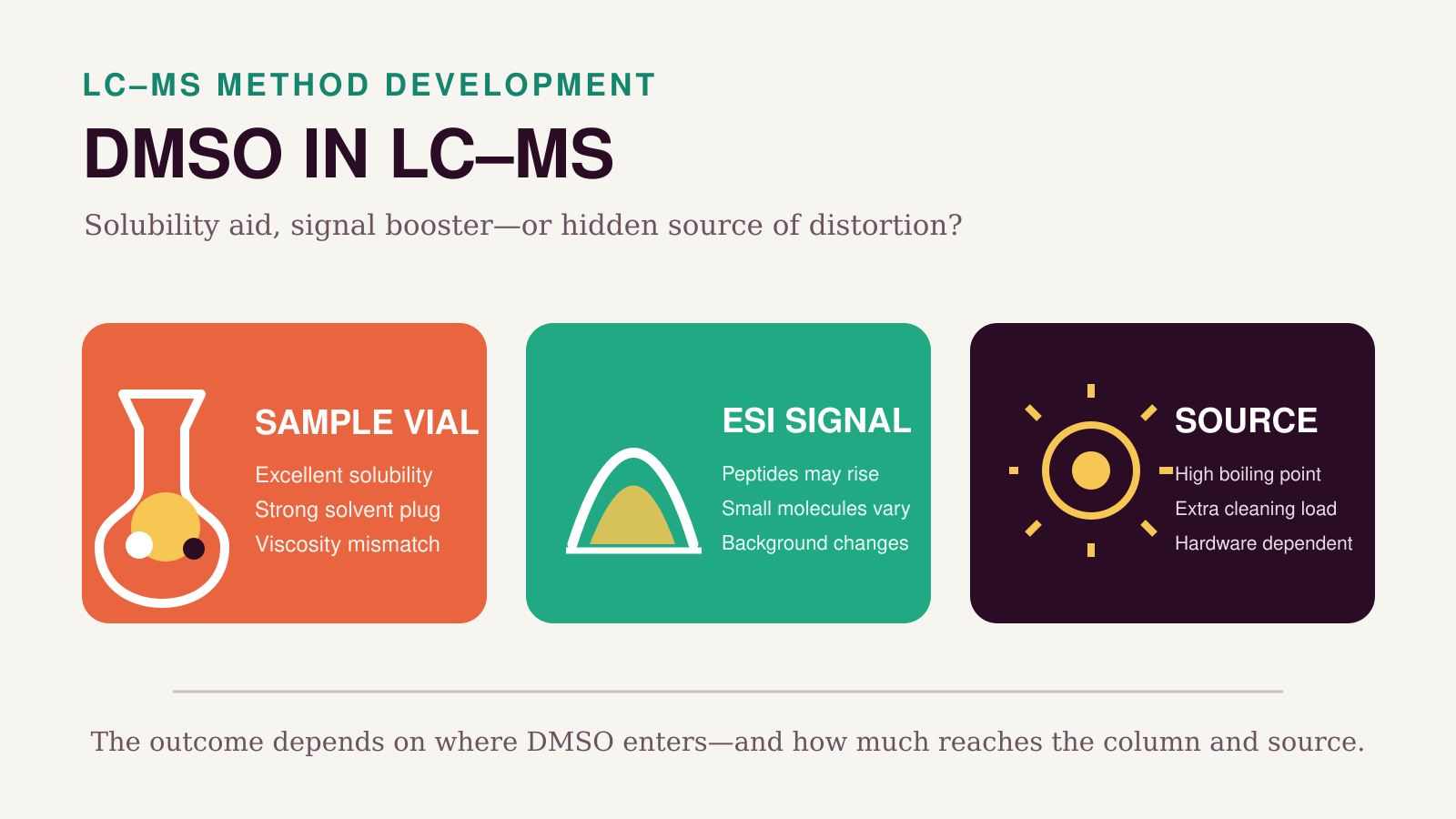
Dimethyl sulfoxide solves compounds that refuse to dissolve in water, acetonitrile or methanol. That makes it indispensable in drug discovery and useful in many analytical workflows. Yet the same physical and chemical properties that make DMSO an exceptional stock solvent can change chromatography, electrospray ionization and source robustness. The outcome depends less on whether DMSO is present than on where it enters the system and at what effective load.

Why DMSO is not just another organic solvent
DMSO is a highly polar, aprotic liquid that is miscible with water and many organic solvents. It dissolves a remarkably broad range of compounds, which is why screening libraries are commonly stored as concentrated DMSO stocks. The analytical benefit is obvious: a compound must be in solution before it can be injected reproducibly.
Its LC–MS behavior is less ordinary. DMSO boils at approximately 189 °C and has low vapor pressure compared with acetonitrile or methanol.1 It is also appreciably more viscous than the common LC modifiers. These properties can slow droplet desolvation and create a viscosity discontinuity when a DMSO-rich sample plug meets the mobile phase. At the same time, DMSO changes droplet surface properties and gas-phase proton-transfer chemistry. The final response is therefore analyte-, source- and method-dependent.
The first question should always be: where is the DMSO?

| How DMSO is used | Main purpose | Primary analytical risk | Best control |
|---|---|---|---|
| Neat stock, then diluted | Solubilize difficult compounds | Residual DMSO differs among standards, QCs and samples | Matrix-match the final DMSO percentage |
| DMSO-rich injection solvent | Avoid precipitation after dilution | Strong-solvent and viscosity effects at the column inlet | Diluent and injection-volume challenge |
| DMSO in mobile phases A and B | Modify ESI response or charge states | Changed gradient strength, selectivity and source load | Re-optimize gradient; compare response and robustness |
| Post-column DMSO addition | Test or exploit an ionization effect | Dilution and altered spray/desolvation | Matched post-column flow without DMSO |
How DMSO affects the LC separation
1. A DMSO-rich plug can behave as a strong solvent in RP-LC
In reversed-phase LC, early analytes are focused when the injection solvent is similar to or weaker than the initial mobile phase. DMSO has substantial elution strength; in the peptide study by Hahne and colleagues it was described as having an elution strength similar to acetonitrile.2 If a sample containing a high DMSO percentage is injected into a water-rich starting gradient, the analyte can move inside the DMSO zone before the programmed gradient begins to elute it normally.
The chromatographic fingerprints are familiar:
- fronting, broadening, shoulders or split peaks;
- reduced apparent retention for early compounds;
- lower peak height even when area changes little;
- loss of resolution close to the void volume;
- a defect that becomes worse as injection volume increases.
This is not a unique DMSO phenomenon. It is the general sample-solvent mismatch problem, intensified by the composition and viscosity of the plug. Peer-reviewed RP-LC work has shown that solvent-strength and viscosity differences can distort peaks, particularly for weakly retained analytes and larger injections.8, 9
2. DMSO can shift an existing gradient
Adding DMSO to both mobile phases is not chromatographically neutral. Hahne et al. had to adapt the gradient to avoid losing hydrophilic peptides when 5% DMSO was present. They also omitted DMSO from the trap-column loading solvent because including it caused a marked overall loss of peptides in their setup.2 This distinction is crucial: DMSO improved the spray response after separation, yet DMSO during loading impaired retention.
A method transferred from 0% to 5% DMSO should therefore be treated as a changed LC method. Verify retention, selectivity, peak width, equilibration, pressure and recovery; do not evaluate only total MS intensity.
3. HILIC is especially sensitive to the injection diluent
HILIC requires a high-organic initial mobile phase and a water-enriched layer at the polar stationary phase. Sample diluent effects are complex because partitioning, hydrogen bonding and electrostatic interactions can all contribute. A systematic nano- and microflow HILIC study found that solvent mismatch could change peak shape and retention, with larger relative injection volumes making the effect more obvious.10
In an earlier silica-HILIC investigation, neat DMSO produced extremely poor peak shape for the tested polar bases. If DMSO was unavoidable, 75:25 acetonitrile/DMSO gave the best result among the tested DMSO mixtures.11 That ratio is useful evidence—not a universal HILIC recipe. The optimum depends on the analyte, stationary phase, initial mobile phase and injection volume.
DMSO can enhance—or suppress—electrospray response
The strongest evidence for enhancement comes from peptides
The best-known DMSO result is a 2013 bottom-up proteomics study. Adding 5% DMSO to both reversed-phase LC solvents increased median peptide signal approximately threefold on an Orbitrap Elite. The benefit was larger for low-intensity peptides, and the detection limit for peptide and protein identification improved by as much as tenfold. Across seven LC–MS/MS systems in three laboratories, summed peptide signal increased by approximately two- to fourfold.2
The effect was not simply narrower peaks. Median chromatographic peaks were only 1.9 seconds broader during a 210-minute separation, and charge-state coalescence explained only about 10–20% of the signal gain. The authors proposed that DMSO altered electrospray droplet behavior and improved the production of gas-phase peptide ions. Their data also showed a shift toward lower peptide charge states and stronger relative benefits for hydrophilic and acidic peptides.
Earlier work had shown that 5% DMSO could concentrate peptide signal into fewer charge states and increase peptide identifications by roughly 10–25%.3 A later urine screening method used DMSO-containing mobile phase to detect 36 small peptide targets and metabolites after only twofold dilution, with reported LODs of 50–1000 pg/mL.5
A separate Top 3 label-free proteomics evaluation found that the DMSO response increase was not uniform across peptides, yet the normalized protein estimates remained strongly correlated with the non-DMSO workflow. The authors concluded that DMSO could be compatible with that specific quantitative strategy, while also showing why an untargeted intensity gain should not be interpreted as an equal response factor for every peptide.4
Why this does not prove a universal sensitivity benefit
Peptides, intact proteins and small molecules do not respond to electrospray in the same way. DMSO can redistribute peptide charge states, compete in proton-transfer reactions, change droplet evaporation and alter background-ion competition. The direction and magnitude of the effect therefore depend on analyte basicity, size, surface activity, mobile-phase acid, flow rate, source geometry and desolvation conditions.
Context dependence is visible even within protein analysis. In experiments with supercharging reagents, 5% DMSO improved protein signals from formic-acid mobile phases but decreased signals when TFA was present under the tested conditions.6 In another workflow, DMSO did not enhance sensitivity in the authors’ nanoflow setup, although it improved other method characteristics.7
DMSO has also been deliberately used as a suppressing agent in mechanistic ESI experiments with a small-molecule analyte.12 The correct conclusion is not that DMSO suppresses all small molecules, but that a peptide-proteomics result cannot be transferred to a quantitative small-molecule assay without direct evidence.
Background ions, carryover and source robustness
DMSO changes the spectrum even when analyte response improves. Hahne et al. observed suppression of common polysiloxane background and the appearance of DMSO-containing ions. They reported an ion at m/z 401.922718 that could serve as a lock mass in their positive-ion setup.2 This can be useful, but it also means that untargeted workflows, background subtraction and inclusion lists may need to be reassessed after DMSO is introduced.
Source robustness deserves equal attention. DMSO’s high boiling point and low volatility increase the desolvation burden relative to ACN or methanol. A Thermo Fisher technical discussion accompanying a proteomics article warned that sustained DMSO use can increase the need for non-routine cleaning and specifically discouraged the additive on the Q Exactive series in that context.13 This is operational guidance, not a cross-platform controlled trial, but it is strong enough that instrument-specific advice should be checked before long sequences.
Do not assume that DMSO itself always increases analyte carryover. In the 2013 peptide study, adding DMSO to the LC solvents actually reduced column carryover.2 Carryover and source fouling are different phenomena: DMSO may improve analyte solubility and wash a sticky compound from the LC path while simultaneously increasing the solvent load that must be removed in the source.
Compatibility also extends beyond the source. Agilent cautions that DMSO can swell PEEK components in certain regeneration procedures.14 Concentration, exposure time and hardware materials matter, so consult the LC and column documentation before using high DMSO percentages as a mobile phase or wash solvent.
A practical experiment to define the acceptable DMSO load
There is no scientifically defensible universal statement that every LC–MS sample must contain less than 0.1%, 1% or 5% DMSO. Those values can be useful starting points, but acceptance must be method-specific. The following challenge isolates the effects efficiently.

- Prepare a DMSO series. A useful initial screen may include 0, 0.1, 0.5, 1, 2 and 5% DMSO in otherwise identical solution. Add higher levels only when the real workflow requires them. Keep matrix, analyte concentration, internal standard, pH and total organic fraction controlled.
- Compare at least two injection volumes. Adjust concentration so that injected analyte mass remains constant. If peak distortion changes with plug volume while analyte mass does not, a solvent effect is more likely than mass overload.
- Record chromatographic metrics. Compare retention time, width, asymmetry or tailing, plate count, resolution and the presence of a solvent-front or breakthrough peak. Inspect the earliest and least retained analytes first.
- Record MS metrics. Compare raw area, peak height, signal-to-noise, internal-standard-normalized response, qualifier ratio or MS/MS spectral quality, background features and response drift through the batch.
- Test the sequence, not only one injection. Include a blank after the high standard, repeated QCs across the batch and a system-suitability injection after the DMSO series. A short-term signal gain is not useful if the source becomes unstable later.
- Match calibrators, QCs and samples. If calibration standards contain 1% DMSO but extracted samples contain 0.1%, the curve may not represent the samples. Equalize the final DMSO concentration wherever practical or validate the difference explicitly.
Calculate the solvent load, not just the vial percentage
A simple and useful descriptor is the volume of neat-DMSO equivalent delivered per injection:
DMSO load = injection volume × DMSO volume fraction
A 10 µL injection containing 2% DMSO delivers 0.20 µL DMSO. A 1 µL injection containing 10% DMSO delivers 0.10 µL. These injections are not automatically equivalent because plug composition also controls mixing and elution strength, but the calculation prevents the misleading habit of discussing percentage without volume.
Troubleshooting DMSO-related LC–MS problems
| Observation | Most likely DMSO-related mechanism | Fast confirmation | First action |
|---|---|---|---|
| Early RP-LC peaks front, broaden or split | Strong DMSO plug and/or viscosity mismatch | Reduce injection volume; reprepare in initial mobile phase | Make an intermediate stock and lower final DMSO |
| HILIC peaks become broad or irregular | Diluent disrupts focusing or the stationary-phase water layer | Compare ACN-rich diluent with identical analyte mass | Increase ACN where solubility permits; reduce volume |
| Signal rises but retention shifts | DMSO is affecting both LC strength and ESI | Add DMSO post-column instead | Re-optimize the gradient or decouple the ionization test |
| Peptide charge states move lower | DMSO-related charge-state coalescence/proton transfer | Compare charge-state distributions | Reassess precursor selection and acquisition settings |
| Small-molecule response falls | Analyte-specific ion suppression or incomplete desolvation | Post-column DMSO step or flow-injection comparison | Lower DMSO; increase dilution; optimize source cautiously |
| Background spectrum changes | DMSO-related ions and altered competition with contaminants | Run mobile-phase blanks with and without DMSO | Update exclusion lists/background filters only after confirmation |
| Response deteriorates during a long batch | Increasing source contamination or desolvation stress | System-suitability trend and source inspection | Stop the sequence; follow manufacturer cleaning guidance |
| Standards and samples show different response | Different final DMSO content or solvent history | Prepare a DMSO-matched control set | Matrix-match or validate the mismatch |
Preferred corrective order
- Dilute the DMSO stock into a compatible intermediate solvent.
- Lower the final DMSO concentration while confirming that the analyte stays dissolved.
- Reduce injection volume and, if possible, increase sample concentration to preserve on-column mass.
- Match standards, QCs and samples for DMSO and total organic content.
- For RP-LC, move the diluent toward the aqueous initial conditions; for HILIC, test a more ACN-rich diluent.
- If DMSO is being considered as an ESI enhancer, test post-column addition before rewriting the LC method.
- Check instrument and flow-path compatibility before sustained high-percentage use.
Bottom line
DMSO has two analytically valuable properties: exceptional solvating power and the ability to alter electrospray behavior. It also has three liabilities: a concentrated plug can disrupt chromatographic focusing, its ionization effect is not universal, and sustained delivery of a high-boiling solvent can increase the burden on the source and flow path.
For routine small-molecule LC–MS, the safest strategy is usually to keep final DMSO as low and consistent as solubility permits, minimize the injection volume and validate both peak shape and normalized response. For peptide LC–MS/MS, deliberate mobile-phase or post-column DMSO addition may deliver a genuine sensitivity advantage—but only after the gradient, charge-state distribution, background and long-sequence robustness have been re-evaluated.
The key distinction is simple: DMSO used to dissolve a sample is not the same analytical intervention as DMSO used to modify electrospray.
References and further reading
- PubChem. Dimethyl Sulfoxide, CID 679. Physical and chemical property record.
- Hahne, H. et al. DMSO Enhances Electrospray Response, Boosting Sensitivity of Proteomic Experiments. Nature Methods 2013, 10, 989–991. DOI: 10.1038/nmeth.2610.
- Meyer, J. G.; Komives, E. A. Charge State Coalescence During Electrospray Ionization Improves Peptide Identification by Tandem Mass Spectrometry. J. Am. Soc. Mass Spectrom. 2012, 23, 1390–1399.
- Strzelecka, D.; Holman, S. W.; Eyers, C. E. Evaluation of DMSO as a Mobile Phase Additive During Top 3 Label-Free Quantitative Proteomics. Int. J. Mass Spectrom. 2015, 391, 157–160.
- Judák, P. et al. DMSO Assisted Electrospray Ionization for the Detection of Small Peptide Hormones in Urine by Dilute-and-Shoot LC-HRMS. J. Am. Soc. Mass Spectrom. 2017, 28, 1657–1665.
- Nshanian, M. et al. Enhancing Sensitivity of LC–MS of Peptides and Proteins Using Supercharging Agents. Int. J. Mass Spectrom. 2018, 427, 157–164.
- Coppieters, G. et al. Combining Direct Urinary Injection with Automated Filtration and LC–MS. 2021. PubMed record.
- Keunchkarian, S.; Reta, M.; Romero, L.; Castells, C. Effect of Sample Solvent on Chromatographic Peak Shape under Reversed-Phase Conditions. J. Chromatogr. A 2006, 1119, 20–28.
- Castells, C. B.; Castells, R. C. Peak Distortion in RP-LC as a Consequence of Viscosity Differences Between Sample Solvent and Mobile Phase. J. Chromatogr. A 1998, 805, 55–61.
- Li, H. et al. A Systematic Investigation of the Effect of Sample Solvent on Peak Shape in Nano- and Microflow HILIC Columns. J. Chromatogr. A 2021, 1655, 462298.
- Grumbach, E. S. et al. Hydrophilic Interaction Chromatography Using Silica Columns for Polar Analytes and Enhanced ESI-MS Sensitivity. Waters application note, 2004.
- Gangl, E. T.; Annan, M. M.; Spooner, N.; Vouros, P. Reduction of Signal Suppression Effects in ESI-MS Using a Nanosplitting Device. Analytical Chemistry 2001, 73, 5635–5644.
- Thermo Fisher Scientific. Using DMSO in Top 3 Label-Free Protein Quantitation. Technical discussion and manufacturer response, 2016.
- Agilent. General Column Regeneration Procedure. Compatibility caution for DMSO and PEEK flow-path components.
Editorial note: The article distinguishes peer-reviewed findings from manufacturer guidance. All three figures are original LC–MS Guide illustrations; no publisher or manufacturer figure was reproduced. The concentration series shown is a proposed validation design, not a universal acceptance limit.
Leave a Reply